

Intraocular Pressure and Glaucoma: What the Numbers on Your Report Actually Mean
A report comes back flagged. Intraocular pressure is elevated. For many people, that sentence raises more questions than it answers.
Intraocular pressure is one of the most clinically important measurements in eye health, yet the number on a report rarely comes with a clear explanation of what it means for you personally. This post covers what eye pressure is, what shifts it, what the readings indicate, and what your options look like when the numbers need attention.
Quick Overview
- Intraocular pressure refers to the pressure created by fluid circulating inside the eye, and keeping it within a healthy range helps protect the optic nerve from damage over time.
- A reading between 10 and 21 mm Hg is generally within the expected range, though this varies depending on the individual and the health of their optic nerve.
- Elevated intraocular pressure does not confirm that glaucoma is present, but it is one of the strongest risk factors for developing it if left unaddressed.
- When pressure remains high, several treatment options exist, from eye drops through to laser treatment and surgery.
Below, we explain what drives eye pressure up, how it is detected early, and what management looks like at each stage.
The Fluid Behind the Pressure
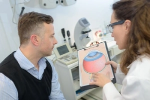 The fluid inside the eye, called aqueous humour, is continuously produced and drains through channels near the base of the iris. It nourishes the internal structures at the front of the eye that have no direct blood supply.
When that drainage works as it should, pressure inside the eye holds steady. When something interferes with drainage, fluid accumulates. The eye cannot expand to accommodate it, so pressure climbs. Sustained elevated eye pressure presses on the optic nerve head, the point where the nerve exits the eye. The optic nerve carries every visual signal your eye generates to the brain. Peripheral vision is typically the first to be affected when nerve fibres are compressed by sustained high IOP, and the cumulative effect of that high pressure on the nerve is what drives glaucoma progression, and that damage, once done, cannot be reversed.
This damage, if it occurs, is irreversible. That is the core reason eye pressure matters.
Reading the Numbers: What Is Considered Normal?
Intraocular pressure IOP is expressed in mm Hg, short for millimetres of mercury. Normal eye pressure for most adults generally sits between 10 and 21 mm Hg, and a healthy intraocular pressure falls comfortably within this band.
But the number is never read in isolation. Your ophthalmologist looks at pressure alongside the appearance of the optic nerve, visual field results, and the patient’s full risk profile. Two people with the same reading can have very different clinical pictures. A reading just above the normal range in an eye with a healthy optic nerve and no other risk factors carries a different clinical meaning than the same reading in someone with a family history of glaucoma and signs of early nerve change.
When pressure climbs above 21 mm Hg
A reading consistently above 21 mm Hg is called ocular hypertension. It is not glaucoma. But it places a person in the category of glaucoma suspects, which means their risk of developing glaucoma over time is meaningfully higher than average. Early treatment at this stage significantly reduces the likelihood of developing glaucoma and can protect the optic nerve before any damage begins.
When pressure sits within the normal range, but damage still occurs
Some patients develop optic nerve damage despite eye pressure sitting comfortably within the normal range. This is called normal tension glaucoma. It tends to reflect either a nerve that is more susceptible to pressure than average or compromised blood flow to the optic nerve head. It is a reminder that pressure is a critical part of the picture, not the entire picture.
Factors That Drive Eye Pressure Higher
Several factors influence how the eye manages fluid, some within your control and some structural.
Drainage pathway changes
Primary open-angle glaucoma is the most common form of the disease. The drainage angle of the eye remains open, but the trabecular meshwork, the fine filtration tissue within that angle, gradually loses its ability to drain fluid efficiently. Pressure rises slowly, silently, and without any symptoms most patients can notice. This is why routine eye exams matter so much; waiting for symptoms to appear means waiting until significant damage has already occurred.
 Risk factors that increase susceptibility
 Risk factors that increase susceptibility
Family history is a significant driver. If a parent or sibling has glaucoma, your own risk is considerably higher. Age increases risk, as does having elevated blood pressure, being significantly short-sighted, having thin corneas, and having diabetes. Glaucoma patients with multiple risk factors are typically monitored more closely and may benefit from earlier treatment even before nerve changes are visible.
Detecting High Eye Pressure Before It Causes Damage
Untreated high eye pressure causes no pain and no visual symptoms you can detect. Eye pressure is measured during a comprehensive eye exam using tonometry, where a small instrument or brief air pulse determines the resistance of the cornea. Because pressure shifts throughout the day, multiple readings across visits build a more reliable picture than any single result. The earlier elevated eye pressure is detected, the better the range of options available.
Managing Elevated Eye Pressure
The goal of treatment is to lower IOP to a level that takes the load off the optic nerve and stops disease progression. The approach depends on where pressure sits, whether nerve damage is already present, and how each patient responds to treatment.
Prescription drops
Prescription eye drops are the usual starting point. Some reduce aqueous humour production; others improve drainage. Consistent daily use is central to keeping pressure controlled, as drops regulate IOP rather than resolve the structural drainage issue.
Laser treatment for the drainage system
Selective laser trabeculoplasty stimulates the trabecular meshwork to improve drainage and lower IOP. Performed in two sessions with no incision required, it delivers meaningful pressure reduction for most patients with primary open-angle glaucoma and can be offered as a first-line option.
When laser and drops are not sufficient
Where other approaches have not achieved the required reduction, surgery creates a new drainage pathway for fluid to leave the eye. It is reserved for advanced or poorly controlled glaucoma and carries a risk profile your ophthalmologist will discuss fully.
Book a Glaucoma Assessment at Mornington Peninsula Eye Clinic
High eye pressure feels like nothing. There is no discomfort, no visual change in most cases, and no signal you can pick up yourself. Routine assessment is the only way to know where your eye pressure sits and whether your optic nerve is being affected.
Mornington Peninsula Eye Clinic provides comprehensive glaucoma assessment and management, including IOP measurement, optic nerve imaging, visual field testing, and access to glaucoma treatment options.
Whether you have received an elevated pressure reading or simply want your eye health assessed, please call us on (03) 9070 3580 or book an appointment online.
Frequently Asked Questions
Is elevated eye pressure always a sign that glaucoma has started?
 Is elevated eye pressure always a sign that glaucoma has started?
No. Elevated intraocular pressure means the eye is producing more fluid than it can drain efficiently, but it does not mean the optic nerve has been damaged yet. Many people with ocular hypertension, meaning pressure consistently above 21 mm Hg, never develop glaucoma. The significance of elevated pressure depends on the full clinical picture, including optic nerve health, risk factors, and corneal thickness. Regular monitoring allows any changes to be caught before vision is affected.
Can eye pressure be lowered without medication?
Certain lifestyle factors are associated with modest reductions in eye pressure for some people. Regular aerobic exercise, limiting caffeine, and avoiding inverted yoga positions have each been associated with modest IOP reductions in some patients. These adjustments contribute to overall eye health but cannot replace clinical management when pressure is clinically elevated.
Does eye pressure need to be checked even if I have no symptoms?
Yes. Elevated intraocular pressure and early glaucoma produce no pain, no visual disturbance, and no warning sign a patient can detect independently. By the time symptoms appear, significant optic nerve damage has typically already occurred. Routine eye pressure checks are the only reliable way to identify elevated IOP before it affects vision, which is why they are a standard part of comprehensive eye examinations regardless of whether any symptoms are present.
Any surgical or invasive procedure carries risks. Before proceeding, seek a second opinion from an appropriately qualified health practitioner.
References
https://www.sciencedirect.com/science/article/pii/S0039625722000388
https://my.clevelandclinic.org/health/symptoms/24552-eye-intraocular-pressure



Leave a Reply
Want to join the discussion?Feel free to contribute!