

Central Retinal Vein Occlusion: Causes, Symptoms and Treatment
If you’ve noticed sudden blurry or distorted vision in one eye, one possible cause is central retinal vein occlusion. This is a blockage in the central retinal vein, the main vein that carries blood away from the retina, which lets blood and fluid back up into the retina. It is a common retinal vascular disorder, and the change in sight can come on within hours or days.
Knowing the early signs matters because the sooner it is checked, the sooner treatment can begin. This guide walks you through what central retinal vein occlusion is, why it happens, how we check for it, and the treatment options that may help protect your vision.
Quick Overview
- Central retinal vein occlusion is a blockage of the central retinal vein, the main vein that drains blood from the retina.
- It usually begins with a blood clot or slowed blood flow, which lets fluid leak into the retina and causes swelling.
- Risk rises with age and with conditions such as high blood pressure, diabetes, high cholesterol and glaucoma.
- Treatment centres on the macular oedema it causes, most often with injections into the eye, and a dilated eye exam confirms the diagnosis.
Below, we begin with what happens inside the eye when the vein becomes blocked and build from there.
What is central retinal vein occlusion?
Central retinal vein occlusion, known as CRVO, is a blockage of the central retinal vein where it leaves the eye near the optic nerve. When blood cannot drain properly, pressure builds in the retinal veins, and fluid leaks into the retina. This causes retinal swelling that can blur your central vision.
The central retinal vein sits alongside the central retinal artery, which brings blood into the eye. A blockage in the vein disrupts the normal blood flow out of the retina. There are two patterns: a milder non-ischaemic type, and a more serious ischaemic type where areas of the retina lose their blood supply. The ischaemic type carries a higher risk of complications and greater vision loss.
What are the symptoms of central retinal vein occlusion?
 The main symptom is blurry or distorted vision in one eye, which may appear suddenly or worsen over several hours or days. Some notice a patchy or dim area in their sight. You might also see floaters, which are small dark spots or lines. The change usually affects only one eye and often happens without pain.
Milder cases may cause few symptoms or vision that comes and goes. More serious cases can lead to significant vision loss. In severe situations where new blood vessels grow, and the eye pressure rises, some experience pain, redness and pressure in the eye. Unlike vision changes that build slowly over months, the shift with this condition is often faster. Any sudden vision loss should be checked without delay.
What causes retinal vein occlusion, and who is at risk?
Most cases begin with a blood clot or slowed blood flow in the vein. This often happens where a stiffened retinal artery crosses and presses on a nearby vein, narrowing it. Age is the leading risk factor, and it is uncommon under 50. The same risk factors that affect the heart and blood vessels also raise the risk here.
The way a blocked vein slows drainage reflects wider blood vessel health. Common risk factors include high blood pressure, diabetes, high cholesterol, glaucoma or elevated intraocular pressure, and smoking. Diabetes also raises the risk of diabetic retinopathy, another condition that can affect the retina. When the condition appears in someone under 40, further blood tests may be arranged to look for a clotting or blood-thickening problem. Your individual risk depends on your health history, so it helps to discuss it with us.
How is retinal vein occlusion diagnosed?
To diagnose retinal vein occlusion, your ophthalmologist starts with a dilated eye exam and checks your visual acuity. Drops widen the pupil so the retina can be viewed closely. Imaging scans then confirm the blockage and measure any swelling in the macula. This helps guide your care.
Two scans are often used. Optical coherence tomography, known as OCT, takes a detailed cross-section of the retina and measures retinal swelling, which helps track macular oedema over time. Fluorescein angiography uses a dye injected into an arm vein and a camera to show which blood vessels are blocked or leaking. A comprehensive eye examination also looks for abnormal new blood vessels and other signs that shape the treatment plan.
What are the treatment options for central retinal vein occlusion?
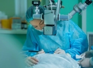 There is no way to unblock the vein itself, so treatment aims to manage the swelling and reduce the risk of complications. The main cause of vision loss is macular oedema, the swelling of the central retina. The current treatment for macular oedema most often involves injections into the eye.
These are known as intravitreal injections, and most use a medication class called anti-VEGF. VEGF stands for vascular endothelial growth factor, a protein that drives leakage and the growth of abnormal blood vessels, so reducing its activity helps ease the macular oedema. Steroid injections are another option, particularly when the response to anti-VEGF is limited.
Laser treatment plays a role in more serious cases. When retinal ischaemia triggers abnormal new blood vessels, a laser can treat the retina to lower the risk of a new blood vessel forming and of neovascular glaucoma, where eye pressure rises. Without care, complications such as retinal detachment can also threaten sight. Alongside eye treatment, managing conditions like high blood pressure and diabetes supports your overall retinal health.
Can you prevent retinal vein occlusion?
You cannot always prevent retinal vein occlusion, but you can lower your risk by looking after your blood vessels. Because the condition shares risk factors with heart disease and stroke, the steps that protect your general health also help your eyes. Regular eye tests support early detection.
To help prevent retinal vein occlusion, it makes sense to control risk factors that damage blood vessels. That means managing high blood pressure, diabetes and cholesterol, staying active, eating well, and not smoking. Managing diabetic eye disease and keeping an eye on glaucoma and eye pressure also matter, since both are linked to the condition. If you have already had an occlusion in one eye, ongoing review helps monitor the other eye.
Getting the Right Care for Retinal Vein Occlusion
A sudden change in your vision is always worth acting on. All central retinal vein occlusions are treated as an urgent clinical priority, which reflects how time-sensitive the condition can be. At Mornington Peninsula Eye Clinic, we assess and monitor retinal vein occlusion as one of the retinal conditions we treat, track macular oedema with detailed imaging, and provide retinal care and surgery when abnormal new blood vessels appear. If you have noticed sudden, blurred or distorted vision in one eye, please call us on (03) 9070 3580 to arrange an assessment.
Frequently Asked Questions
Can vision recover after a central retinal vein occlusion?
Recovery varies from person to person and depends on the type and severity of the blockage. Some regain useful vision, especially with milder cases and treatment of macular oedema, while vision may not return to how it was before. Ischaemic cases tend to have a poorer outlook. Ongoing monitoring helps us adjust care over time.
What is the difference between central and branch retinal vein occlusion?
Central retinal vein occlusion, or CRVO, affects the main central retinal vein and usually influences the whole retina. Branch retinal vein occlusion, or BRVO, affects one of the smaller branch veins, so it involves a smaller area. BRVO is the more common of the two. Both can cause macular oedema and vision changes, and both need assessment.
Is central retinal vein occlusion the same as a central retinal artery occlusion?
No. A vein occlusion blocks blood draining out of the retina, while a central retinal artery occlusion blocks blood flowing in. Artery occlusions often cause more sudden and severe vision loss and are treated as an emergency. Any sudden vision loss should be checked straight away, whichever vessel is involved.
Can retinal vein occlusion affect both eyes?
Retinal vein occlusion usually affects one eye at a time. A smaller number of patients later develop a blockage in the other eye. Having had one occlusion raises the risk to the fellow eye, which is why managing your risk factors and attending review appointments is worthwhile.
How often are eye injections needed for macular oedema?
This differs for each patient and each eye. Because swelling can return as the medicine wears off, these injections are often repeated and then spaced out based on how your retina responds. We use OCT scans to guide the timing, so the plan is tailored rather than fixed.
How quickly should I get my eyes checked after sudden vision loss?
Sudden vision loss should be treated as an ocular emergency, so seek an eye assessment as soon as possible rather than waiting to see if it improves. Prompt review lets us find the cause, check for macular oedema and abnormal blood vessels, and begin any treatment early. Booking an assessment quickly means the cause can be identified without delay.
Any surgical or invasive procedure carries risks. Before proceeding, you should seek a second opinion from an appropriately qualified health practitioner.
References
https://my.clevelandclinic.org/health/diseases/14206-retinal-vein-occlusion-rvo
https://www.nejm.org/doi/full/10.1056/NEJMicm2518603



Leave a Reply
Want to join the discussion?Feel free to contribute!