

Retinal Tear Surgery: How a Tear Is Sealed Before It Spreads
Flashes at the edge of your vision, or floaters that arrived suddenly, are usually harmless. Occasionally, an examination finds a tear in the retina, and the question becomes what happens next and how soon.
Retinal tear surgery is one of several ways a tear at the back of the eye is dealt with, and which one suits you depends on where your tear sits and what caused it. Below you will find why tears form, how the seal is created, and what the alternatives are when a laser is not suitable.
Quick Overview
- A retinal tear happens when the vitreous gel shrinks with age and pulls hard enough to open a break in the retina.
- Sealing the break with small burns is what seals it, since those burns create scar tissue as they heal, pinning the retina down.
- Laser photocoagulation is the usual choice, with cryotherapy and pneumatic retinopexy used in particular situations.
- A tear treated early rarely progresses, while an untreated tear can lead to retinal detachment and permanent vision loss.
The sections below cover how tears form, the warning signs, how each treatment works, and what the recovery involves.
What happens when the vitreous pulls away
Light entering your eye lands on the retina, a thin layer of tissue at the back that turns it into signals your brain can read. Occupying the space in front is a clear gel-like substance called the vitreous, which presses against that layer.
Age shrinks that gel. When the vitreous separates, it normally comes away without incident, and the floaters that show up during the process are harmless. Where the gel stays firmly attached at one point, the pulling can open a break in the tissue beneath.
Multiple factors make this more likely. Among the risk factors, short-sightedness is the most common, along with a family history of retinal conditions, previous eye surgery, eye injury and diabetic eye disease. Age contributes too, since the vitreous changes most in the decades after fifty.
The warning signs, and why they matter
A retinal tear causes no pain. In the early stages, it produces increased floaters, often a sudden increase in them, flashes of light at the edge of your vision, or blurred vision where the tear causes bleeding into the gel.
Those symptoms deserve an examination within days. Left untreated, a tear allows fluid to seep beneath the retina and lift it away from the wall of the eye, which is a detached retina. Reduced vision follows. Once the retina has come away, recovering lost vision is no longer certain, and permanent loss of vision becomes a real possibility.
A shadow or curtain moving across your field of view means the process has already started. That is a medical emergency, and it needs an emergency department or triple zero on the same day.
How laser photocoagulation seals a tear
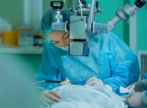 Retinal laser works on a simple principle. Retinal tissue absorbs the light the laser emits and turns it into heat. Rows of small burns are placed in a ring around the break, and the healing process converts each one into a permanent scar that bonds the retina to the layer underneath.
The purpose is a firm adhesion around the break that stops fluid spreading underneath. You sit at a slit lamp for laser surgery of this kind, with a special lens resting against the affected eye. For breaks further out towards the retinal edge, a headset is used instead.
Anaesthetic eye drops numb the surface beforehand. You will see the laser beam as bright flashes during treatment. There is no incision and nothing to stitch, and a single sitting covers it for most patients.
When laser is not the right tool
Lasers are not suitable for every tear. Where a tear sits too far forward to reach, or where bleeding in the vitreous blocks the view, freezing treatment does the same job from outside the eye. A probe applied to the surface of the eye creates the same scar and the same seal.
Cryotherapy tends to be less comfortable than laser and is usually done under local anaesthetic. Both approaches treat retinal tears to the same end. The choice between them turns on several factors specific to your eye, chiefly the position of the tear and what can be seen at the examination.
A third option applies where fluid has begun to collect beneath the retina. In pneumatic retinopexy, the tear is frozen, and a gas bubble is injected into the eye to press the retina back into position. Where a detachment is larger, retinal surgery goes further again. Silicone oil during a vitrectomy is one route, and stitching a scleral buckle around the outside of the eyeball is another.
Recovery and what to avoid
You go home the same day. Temporary blurry vision lasts a few hours while the dilating drops wear off, so arrange a driver instead of planning to get yourself home.
The seal strengthens as the scar tissue matures, so heavy lifting and strenuous activities go on hold briefly. How long that lasts is something your eye surgeon will tell you, since treatment depends on various factors, chiefly the size and position of the tear.
Follow-up appointments check that the seal is holding, and they include a look at the rest of the retina. Protecting your overall eye health means attending those reviews even when your vision feels normal.
 Talk to Mornington Peninsula Eye Clinic about your symptoms
 Talk to Mornington Peninsula Eye Clinic about your symptoms
Most flashes and floaters turn out to be nothing, and telling those apart from the urgent ones takes a dilated examination of the peripheral retina. Our retinal ophthalmologist looks at the whole retina, seals tears with a laser during the same visit where that is appropriate, and sets out the potential risks and treatment options before you decide anything. To arrange an appointment, please call us on (03) 9070 3580.
Frequently Asked Questions
Will I need time off work after retinal tear surgery?
It depends on what your work involves. Desk-based work is generally manageable once your vision has cleared, while physical work involving lifting or straining needs longer. Your surgeon will give you a timeframe on the day.
Is retinal tear treatment covered by Medicare?
With a valid referral, a rebate applies through Medicare. What you pay beyond that varies with the procedure, and you will be given the figures for your own situation before anything proceeds.
Can both eyes develop retinal tears?
Yes, and having had one raises the likelihood in the other eye. The vitreous changes in both eyes over time, so new flashes or floaters on the other side are worth reporting promptly.
Do floaters disappear once a retinal tear is sealed?
Not usually. What you are seeing are shadows thrown by the gel itself, and closing a break does nothing to remove them, though many become less noticeable over time. A sudden shower of new ones afterwards is a separate matter and needs reporting.
Can a sealed retinal tear open up again?
The treated area holds. What can happen is a fresh tear forming in another part of the retina, particularly while the vitreous is still pulling away, which is the reason for follow-up and not a sign the first treatment failed.
Any surgical or invasive procedure carries risks. Before proceeding, you should seek a second opinion from an appropriately qualified health practitioner.
References
https://www.betterhealth.vic.gov.au/health/conditionsandtreatments/retinal-detachment
https://www.mayoclinic.org/diseases-conditions/retinal-detachment/diagnosis-treatment/drc-20351348



Leave a Reply
Want to join the discussion?Feel free to contribute!